Most people are probably aware that shark skeletons are largely composed of cartilage rather than bone. When you hear cartilage you may be thinking of the elastic cartilage of your ear or the hyaline cartilage that forms the tip of your nose. Shark cartilage, however, is a special type of cartilage that is not found elsewhere and is especially strong. Nonetheless, its density is still lower than bone which makes it hard to see in CT scans.
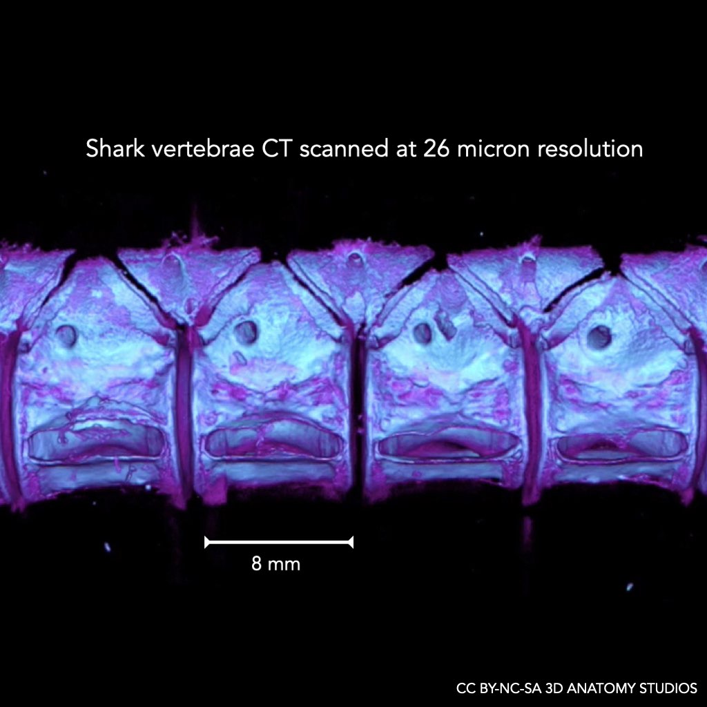
One exception to this is the vertebrae of sharks, which have some of the densest cartilage in the body, potentially because of the extra strength needed to stiffen the axial column. In 2021 we began an initiative to CT scan animals commonly used in anatomy courses and make them freely available online and because of the higher density of shark vertebrae, we suspected that we would be able to resolve many of the anatomical features. So we dissected out a section of vertebral column from a shark dissection specimen and scanned these in a microCT scanner at 26 micron resolution.
This image shows the result of that scan in remarkable detail. The four barrel-like structures on the lower half of the vertebral column are the centra. Above these centra is where the spinal cord runs. The spinal cord is protected by surrounding upper dorsal intercalary plates (inverted triangles) and lower neural arches (non-inverted triangles). Both of these plates have holes in each segment so that the dorsal (upper) and ventral (lower) roots can branch off from the spinal cord and go to the structures of the body that they innervate.
This CT scan of the shark vertebrae is freely available for download here.
Created by: Aaron Olsen, PhD
Software used: 3D Slicer
License: CC BY-NC-SA 3D Anatomy Studios