If you think of a shark tooth, the first image to come to mind is probably the large serrated dagger-shaped tooth of a white shark (commonly referred to as a “great white shark”). However, just as there are many species of sharks, there are also many different sizes and shapes of shark teeth, depending on the size of the shark and what they eat.
The spiny dogfish shark is a shark that is commonly dissected in high school and college anatomy courses. Because spiny dogfish sharks mostly eat small, hard-shelled prey they have teeth that are specialized for grasping, cutting and crushing rather than piercing. However, even if students get the opportunity to dissect a specimen, they usually won’t even see the shark’s teeth. This is in part because the shark’s mouth is preserved closed and it’s very difficult to open the mouth to see the teeth. But it’s also because the teeth are so small, just 3 mm long in the largest dissection specimens.
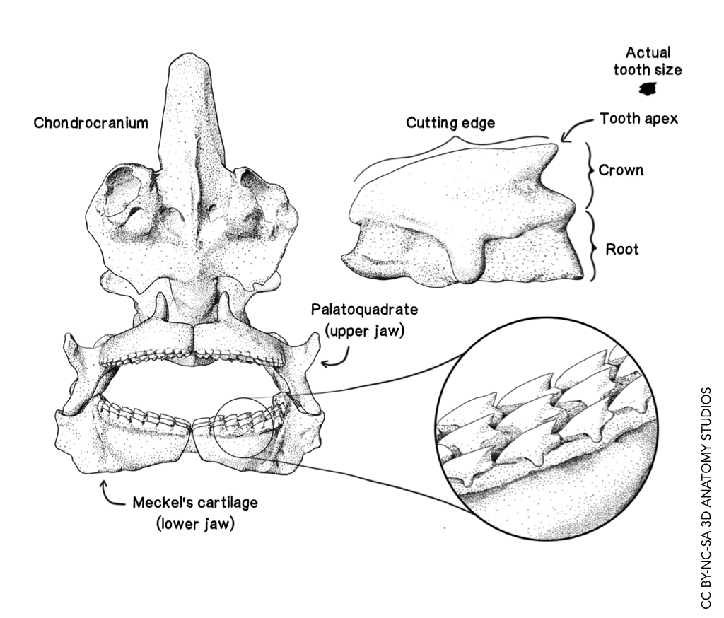
The first 3D digital model that we created was of a spiny dogfish shark tooth, based on images of actual dogfish shark teeth viewed through a dissecting microscope. This enabled us to 3D print the tooth 20x larger than life size so that students can actually feel the cusp, cutting edge, and other key features of the tooth. However, we also needed a visualization that would explain these features of the tooth, how the teeth are arranged in the jaw, and how the jaws are positioned within the shark skull.
To accomplish this, 3D Anatomy Studios member Michael Fath created this illustration of the dogfish shark tooth in context. The focal illustration of the individual tooth labels its key components. The focal illustration of multiple teeth aligned within the jaw shows how the individual teeth are aligned to create a nearly continuous cutting surface and a series of “barbs” that hold prey in place. And the illustration of the skull and jaws shows the size of the teeth relative to the size of the jaws and how multiple rows of teeth on the upper and lower jaws create an impressive biting battery.
Illustrated by: Michael Fath, MS
3D model reference material by: Aaron Olsen, PhD
Software used: Adobe Illustrator, Blender for creating 3D model reference material
License: CC BY-NC-SA 3D Anatomy Studios


